References used in this project
Albert, A.M. and Maples, W.R. (1995). Stages of epiphyseal union for thoracic and lumbar vertebral centra as a method of age determination for teenage and young adult skeletons. Journal of Forensic Sciences 40: 623–633.
Anderson, M., Green, W.T. and Messner, M.B. (1963). Growth and the predictions of growth in the lower extremities. Journal of Bone and Joint Surgery 45A: 1–14.
Anderson, M., Messner, M.B. and Green, W.T. (1964). Distribution of lengths of the normal femur and tibia from one to eighteen years of age. Journal of Bone and Joint Surgery 46A: 1197–1202.
Ashley, G.T. (1954). The morphological and pathologi¬cal significance of synostosis at the manubrio-sternal joint. Thorax 9: 159–166.
Ashley, G.T. (1956). The relationship between the pat¬tern of ossification and the definitive shape of the meso¬sternum in man. Journal of Anatomy 90: 87–105.
Birkner R. (1978). Normal Radiographic Patterns and Vari¬ances of the Human Skeleton – an X-ray Atlas of Adults and Children. Baltimore, Munich: Urban and Schwarzenberg.
Brodeur, A.E., Silberstein, M.J. and Graviss, E.R. (1981). Radiology of the Pediatric Elbow. Boston, MA: G.K. Hall.
Caffey, J. and Madell, S.H. (1956). Ossification of the pubic bones at birth. Radiology 67: 346–350.
Cardoso, H.F. (2008). Age estimation of adolescent and young adult male and female skeletons II, epiphyseal union at the upper limb and scapular girdle in a mod¬ern portuguese skeletal sample. American Journal of Phys¬ical Anthropology 137: 97–105.
Cardoso, H.F. (2010). The chronology of epiphyseal union in the hand and foot from dry bone observations. International Journal of Osteoarchaeology 20: 737–746.
Cardoso, H., Campanacho, V., Gomes, J. and Marinho, L. (2013). Timing of fusion of the ischiopubic ramus from dry bone observations. HOMO-Journal of Compara¬tive Human Biology 64: 454–462.
Doherty, B.J. and Heggeness, M.H. (1994). The quanti¬tative anatomy of the atlas. Spine 19: 2497–2500.
Elgenmark, O. (1946). The normal development of the ossific centres during infancy and childhood. Acta Pedi¬atrica Scandinavica 33(Suppl. 1).
Fawcett, E. (1907). On the completion of ossifica¬tion of the human sacrum. Anatomischer Anzeiger 30: 414–421.
Flecker, H. (1942). Time of appearance and fusion of ossification centres as observed by roentgenographic methods. American Journal of Roentgenology 47: 97–159.
Gardner, E. and Gray, D.J. (1970). The prenatal devel¬opment of the human femur. American Journal of Anat¬omy 129: 121–140.
Greulich, W.W. and Pyle, S.I. (1959). Radiographic Atlas of Skeletal Development of the Hand and Wrist. Stanford: Stanford University Press.
Hansman, C.E. (1962). Appearance and fusion of ossifi¬cation centres in the human skeleton. American Journal of Roentgenology 88: 476–482.
Jenkins, F.A. (1969). The evolution and development of the dens of the mammalian axis. Anatomical Record 164: 173–184.
Love, S.M., Ganey, T. and Ogden, J.A. (1990). Postnatal epiphyseal development: the distal tibia and fibula. Jour¬nal of Pediatric Orthopaedics 10: 298–305.
MacLaughlin, S.M. (1990a). Epiphyseal fusion at the sternal end of the clavicle in a modern Portuguese skel¬etal sample. Antropologia Portuguesa 8: 59–68.
Maclean, S.J., Black, S.M. and Cunningham, C.A. (2014). The developing juvenile ischium: macro‐radio¬graphic insights. Clinical Anatomy 27: 906–914.
McCarthy, S.M. and Ogden, J.A. (1982a). Radiology of postnatal skeletal development. V Distal humerus. Skel¬etal Radiology 7: 239–249.
McCarthy, S.M. and Ogden, J.A. (1982b). Radiology of postnatal development. VI. Elbow joint. Skeletal Radiol¬ogy 9: 17–26.
McKern, T.W. and Stewart, T.D. (1957). Skeletal Age Changes in Young American Males, Analysed from the Stand¬point of Age Identification Technical Report EP-45. Natick, MA: Headquarters Quartermaster Research and Devel¬opment Command.
Milgram, J.W. and Lyne, E.D. (1975). Epiphysiolysis of the proximal femur in very young children. Clinical Orthopaedics and Related Research 110: 146–153.
Moss, M.L. and Noback, C.R. (1958). A longitudinal study of digital epiphyseal fusion in adolescence. Ana¬tomical Record 131: 19–32.
Noback, C.R., Moss, M.L. and Leszczynska, E. (1960). Digital epiphyseal fusion of the hand in adolescence: a longitudinal study. American Journal of Physical Anthropol¬ogy 18: 13–16.
Ogden, J.A., Beali, J.K., Conlogue, G.J. and Light, T.R. (1981). Radiology of postnatal development. IV. Distal radius and ulna. Skeletal Radiology 6: 255–266.
Ogden, J.A. and Phillips, S.B. (1983). Radiology of post¬natal skeletal development. VII. The scapula. Skeletal Radiology 9: 157–169.
O’Rahilly, R., Gardner, E. and Gray, D.J. (1959). The skeletal development of the hand. Clinical Orthopedics and Related Research 13: 42–51.
Osborne, D.R., Effmann, E.L., Broda, K. and Harrelson, J. (1980). Development of the upper end of the femur with special reference to its internal architecture. Radi¬ology 137: 71–76.
Patcas, R., Signorelli, L., Peltomaki, T. and Schatzle, M. (2013). Is the use of the cervical vertebrae maturation method justified to determine skeletal age? A compari¬son of radiation dose of two strategies for skeletal age estimation. European Journal of Orthodontics 35: 604–609.
Piatt, J.H., Jr. and Grissom, L.E. (2011). Developmental anatomy of the atlas and axis in childhood by computed tomography: clinical article. Journal of Neurosurgery Pedi¬atrics 8: 235–243.
Prakash, S., Chopra, S.R.K. and Jit, I. (1979). Ossifica¬tion of the human patella. Journal of the Anatomical Soci¬ety of India 28: 78–83.
Ríos, L. and Cardoso, H.F.V. (2009). Age estimation from stages of union of the vertebral epiphyses of the ribs. American Journal of Physical Anthropology 140: 265–274.
Scheuer, J.L. and MacLaughlin-Black, S.M. (1994). Age estimation from the pars basilaris of the fetal and juvenile occipital bone. International Journal of Osteoar¬chaeology 4: 377–380.
Scranton, P.E., McMaster, J.H. and Kelly, E. (1976). Dynamic fibular function. A new concept. Clinical Ortho¬pedics and Related Research 118: 76–81.
Shapiro, R. and Robinson, F. (1976). Anomalies of the cranio-vertebral border. American Journal of Roentgenol¬ogy 126: 1063–1068.
Shirley, N.R. and Jantz, R.L. (2011). Spheno-occipital synchondrosis fusion in modern Americans. Journal of Forensic Sciences 56: 580–585.
Stevenson, P.H. (1924). Age order of epiphyseal union in man. American Journal of Physical Anthropology 7: 53–93.
Tillmann, B. and Lorenz, R. (1978). The stress at the human atlanto-occipital joint. Anatomy and Embryology 153: 269–277.
Torgersen, J. (1950). A roentgenological study of the metopic suture. Acta Radiologica 33: 1–11.
Walker, G.E. and Kowalski, C.J. (1972). On the growth of the mandible. American Journal of Physical Anthropology 36: 111–118.
Weaver, D.S. (1979). Application of the likelihood ratio test to age estimation using the infant and child tem¬poral bone. American Journal of Physical Anthropology 50: 263–270.
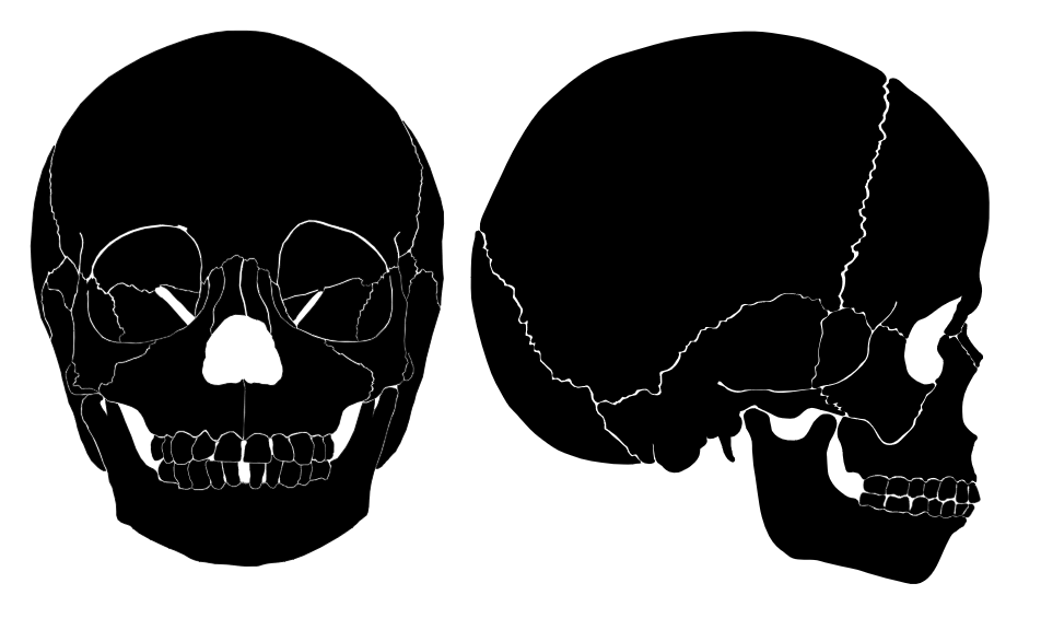
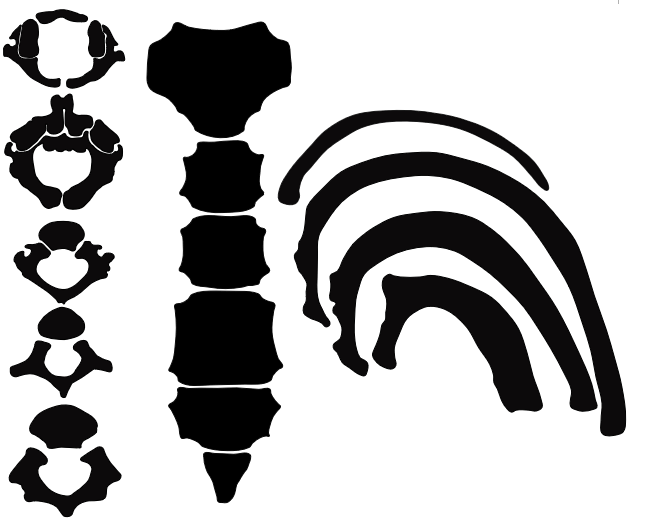
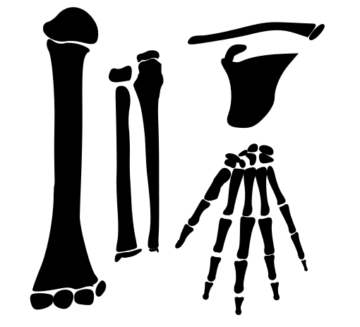
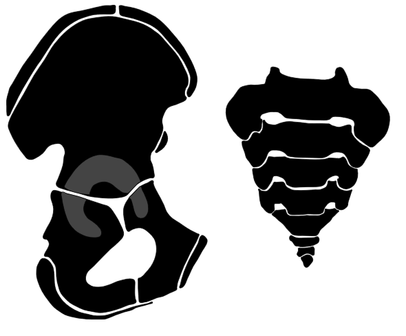
Jump to a region