Lower Limbs Epiphyseal fusion in the legs and feet

image/svg+xml

Click on any of the bones of the skull to take you to that bone's age estimator.

Bones of the lower limbs

The bones of the thorax used for juvenile aging in this project are:

  1. Compare the bone/element you have to the quick description of aging methods provided for that specific bone.
  2. Select the level of fusion or development for the bone provided in the Age Estimator box to receive an age estimation.


Femur
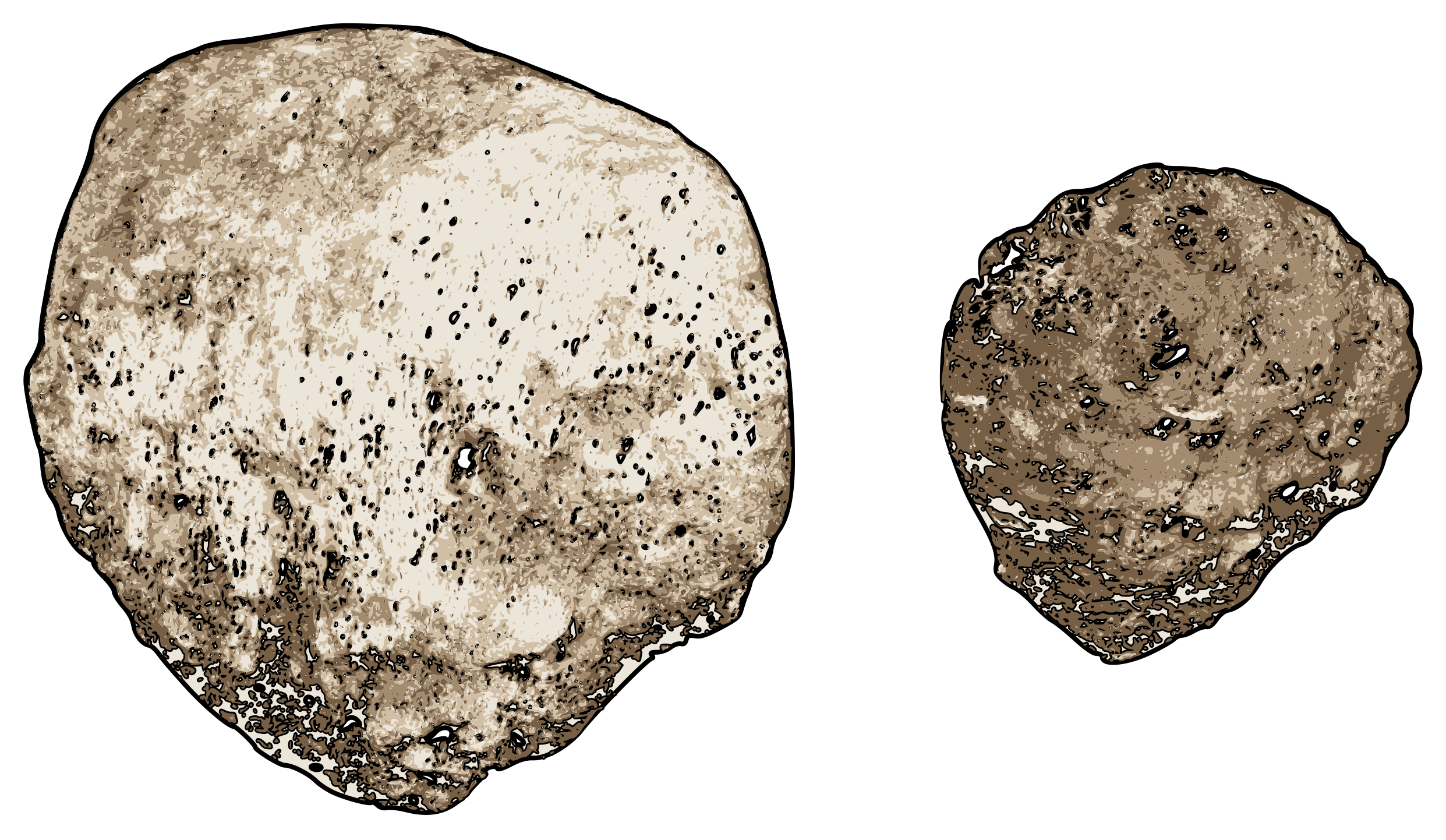
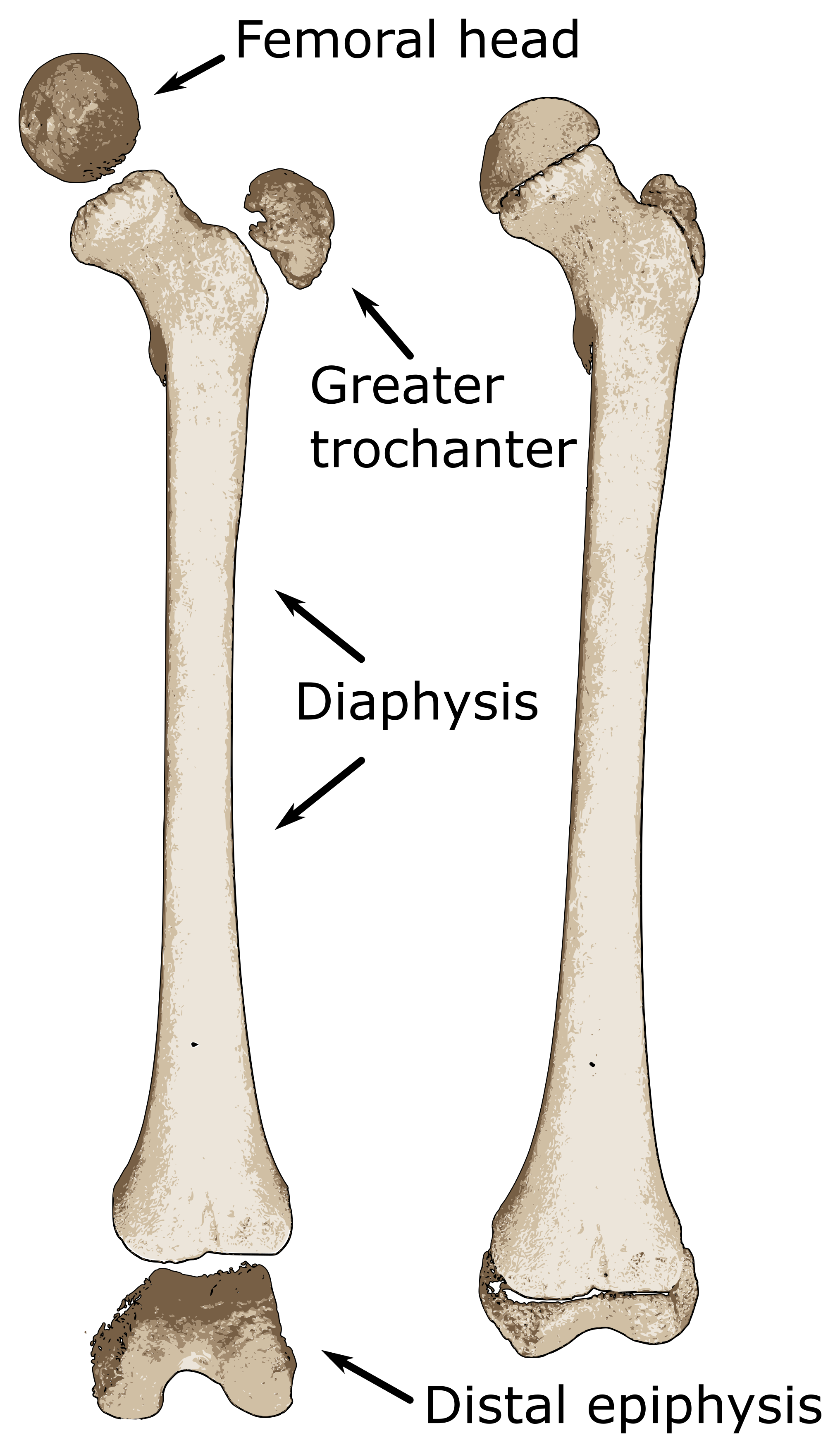
Juvenile femur with unfused epiphyses.
Femur

Femur

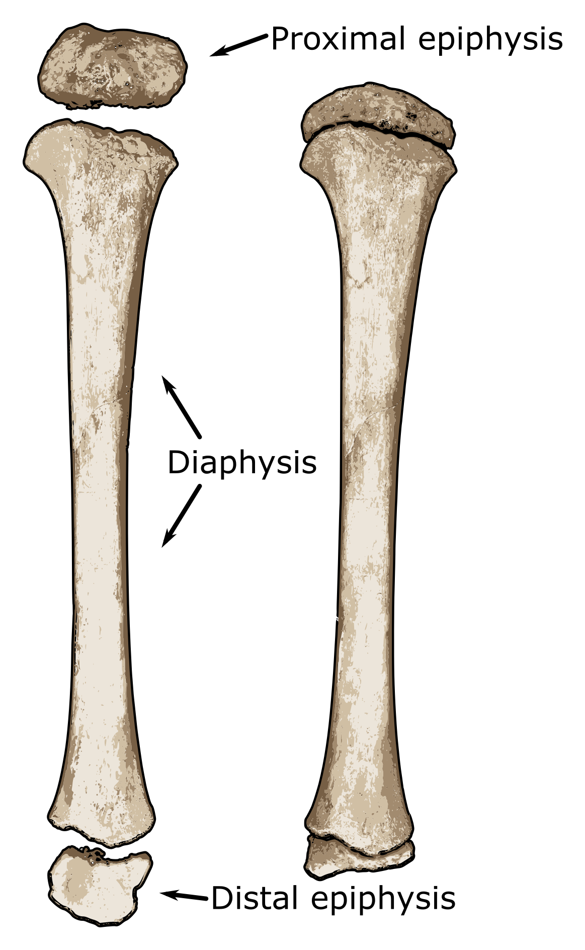
Unlike many other bones, the femur is represented by both the diaphysis and the distal epiphysis at birth (Gardner and Gray 1970). Within the first year of life, the ossification center for the femoral head appears while between 2 and 5 years, the ossification center for the greater trochanter commences (Milgram and Lyne 1975). It is not until 7 to 12 years of age when the ossification center for the lesser trochanter appears.

Fusion times of the femur are variable and somewhat sex-specific. The femoral head fuses between 12 and 16 years old in females and between 14 and 19 years in males. The same pattern goes for the greater trochanter: 14-16 females; 16-18 males (Milgram and Lyne 1975; Scheuer and Black 2004). The lesser trochanter appears to fuse evenly between males and females from 16 to 17 years old.

The last epiphyses to fuse are the distal epiphyses. Again, there appear to be sex-related differences: 14-18 in females; 16-20 in males .


Femoral head


Estimated Age Range:

Greater trochanter


Estimated Age Range:

Lesser trochanter


Estimated Age Range:

Distal epiphysis - condyles


Estimated Age Range:


Patellae
Patellae from an older child (left) and one from a younger child (right).
Patella

Patella

Since the patella has no separate ossification centers that fuse to it, it’s best to look at the overall morphology. The patella appears between 1½ and 3½ years. By 4-5 years old, it is still very coarse and tough, but it does begin to take shape. By puberty, it has essentially taken on its adult form (Prakash et al. 1979).


Patella Presentation


Estimated Age Range:


Tibia
Juvenile tibia showing unfused proximal and distal epiphyses.
Tibia

Tibia

Similar to the femur, the tibia may be represented by two ossification centers at birth: the diaphysis and the proximal epiphysis. If not present at birth, they usually appear within the next few weeks (Scheuer and Black 2004).

Within the next/first 10 months of life, the secondary centers for the distal epiphyses begin to ossify and between 3 and 5 years, and the medial malleolus commences ossification. The distal epiphyses start fusing to the diaphysis between 14-16 years (females) and 15-18 years (males) (Love et al. 1990).

Fusion of the proximal epiphysis comes a little bit later in most cases, between 13 and 17 years old in females and 15 and 19 in males.


Proximal epiphysis


Estimated Age Range:

Distal epiphysis


Estimated Age Range:


Fibula
Juvenile fibula with the distal epiphysis. Note: proximal (head of the fibula) epiphysis is absent due to preservation of the remains.
Fibula

Fibula

Compared to other bones, the commencement, timing, and order of ossification centers for the fibula is comparatively normal. At birth, the individual is only represented by the diaphysis and no epiphyses, unlike it is with the femur and the tibia (Scranton et al. 1976).

Between 10 and 22 months, the distal epiphysis begins its ossification process. During the 4th and 5th years, the proximal ossification centers begin ossification (Love et al. 1990). Like with many other bones, it appears that females grow and develop their skeletal system much more quickly than males. The distal epiphysis fuses between 12 and 15 years (in females) and 15-18 in males (Scheuer and Black 2014).

Lastly, the proximal epiphysis fuses to the fibula, making up the ‘head of the fibula’. In females, this occurs between 12 and 17 years while for males, it fuses between 15 and 20 years on average.


Proximal epiphysis - head


Estimated Age Range:

Distal epiphysis - lateral malleolus


Estimated Age Range:


Tarsals
Feet

Feet


Tarsals

Like the carpals in the wrists, tarsals in the ankle make up an incredibly complex joint. To accompany that complex joint is the complicated way that the bones ossified. Similar to the way that the carpals are dealt with on this site, only the appearance of tarsals will be mentioned due to their complex timetable (Cardoso 2010). (Note: the absence of one tarsal does not mean that the particular tarsal was never ossified, but that it may be lost or degraded due to poor preservation.)


Presence of tarsals:


Estimated Age Range:


Metatarsals
The bones on the left show the first metatarsal and is base epiphysis while the skinnier and longer bones on the right represent metatarsals 2-5 and their head epiphyses.

Metatarsals

Like in the metacarpals in the hand, ossification for the metatarsals on digits 2-5 is different than for the first metacarpal. All metatarsal bodies are present at birth, however the base of the first metatarsal doesn’t appear until the individual is 2-3 years old, with fusion not complete until 13-15 years (female) or 16-18 years (male) (Davies et al. 2013).

For the metatarsals of digits 2-5, the epiphyseal heads don’t appear until 2-3 years of age similar to the base of the first metatarsal. The heads fuse to the rest of the metatarsals somewhat earlier than does the base of the first phalanx though. Fusion appears to be regulated to some extent by the individual’s sex, but fusion times occur around 11-13 years old for females, and 14-16 for males (Scheuer and Black 2004).


Fusion of base to 1st metatarsal:


Estimated Age Range:

Fusion of heads to metatarsals 2-5


Estimated Age Range:


Pedal phalanges
The bones on the left show the proximal and distal pedal phalanges and their base epiphyses while the bones on the right represent the proximal, middle, and distal pedal phalanges (2-5) and their base epiphyses.

Pedal phalanges

The appearance and fusion of pedal phalanges is very similar to manual phalanges. All of the phalanges are present at birth, but have somewhat different appearance and fusion times.

Bases for the proximal phalanges appear around age 1-2 years old and fuse between 13 and 15 years old (females) and 16-18 years old (males). The appearance of epiphyseal bases for the middle and distal phalanges are only slightly different. Middle phalangeal bases start to ossify around 1-2 years while bases for distal phalanges ossify around 2-3 years (Schuler-Ellis and Lazar 1984).

However, fusion times of the bases to both the middle and distal phalanges take place around the same time: 11-13 years for females and 14-16 for males.


Fusion of bases to proximal phalanges


Estimated Age Range:

Fusion of bases to intermediate and distal phalanges


Estimated Age Range: